
Chronic Eye Conditions After 65: Expert Management Tips
Your Vision, Your Future: Taking Control Today
Managing glaucoma and AMD after 65 requires commitment, but the rewards are substantial. With today’s advanced treatments and the remarkable research breakthroughs on the horizon, maintaining functional vision throughout your senior years is not just possible—it’s achievable.
Remember these key takeaways:
- Regular comprehensive eye exams remain your most powerful tool for preserving vision
- Early detection and prompt treatment offer the best outcomes for both conditions
- Daily medication adherence makes the critical difference between stable vision and progression
- Lifestyle modifications complement medical treatments and significantly improve outcomes
- Low vision aids and home adaptations maintain independence even with vision changes
- Support resources help you navigate both emotional and practical challenges
- Ongoing# Managing Chronic Eye Conditions Like Glaucoma and AMD After 65: Your Complete Guide to Preserving Vision
After turning 65, maintaining healthy vision becomes more critical than ever. Chronic eye conditions like glaucoma and age-related macular degeneration (AMD) affect millions of seniors, but with proper management and early intervention, you can preserve your independence and quality of life. This guide walks you through everything you need to know about living well with these conditions.
Understanding Glaucoma and Macular Degeneration in Seniors
What Makes Age 65 a Turning Point for Eye Health
Your eyes undergo significant changes as you reach your mid-60s. The risk of developing serious eye diseases increases dramatically during this time. Studies show that nearly one in three Americans over 65 experiences some form of vision loss, making this decade crucial for proactive eye care.
Both glaucoma and AMD share common risk factors related to aging, including changes in blood vessel health, decreased cellular repair mechanisms, and accumulated oxidative stress over decades. Understanding these conditions early helps you recognize warning signs and seek appropriate care before vision loss occurs.
Glaucoma: The Silent Thief of Sight
Glaucoma describes a group of eye diseases that damage the optic nerve, typically caused by elevated pressure inside the eye. This condition earned its nickname as the “silent thief of sight” because it progresses gradually, often without noticeable symptoms, until significant vision loss occurs.
The most common form, open-angle glaucoma, accounts for about 90% of cases. In this type, the drainage system in your eye becomes less efficient over time, causing fluid buildup and increased pressure that damages the optic nerve. Without treatment, glaucoma leads to irreversible peripheral vision loss and, eventually, complete blindness.
Key statistics about glaucoma:
- Affects approximately 4.22 million Americans (2022 data), with nearly half unaware they have it
- Among those 65 and older, 5.2% have glaucoma
- Glaucoma is the leading cause of irreversible blindness worldwide
- Adults over 60 face a significantly higher risk, especially African Americans (3 times more likely to have vision-affecting glaucoma than white individuals) and Hispanics
- Family history increases your risk substantially
Age-Related Macular Degeneration: Losing Central Vision
AMD affects the macula, the central part of your retina responsible for sharp, detailed vision needed for reading, recognizing faces, and driving. Unlike glaucoma, which impacts peripheral vision first, AMD compromises your central vision while typically preserving side vision.
Two types of AMD exist:
Dry AMD represents about 90% of cases. This form develops slowly as tiny yellow deposits called drusen accumulate under the macula, causing it to thin and deteriorate gradually. While currently no cure exists for dry AMD, recent treatment breakthroughs in 2025 offer new hope.
Wet AMD accounts for the remaining 10% but causes more severe, rapid vision loss. In wet AMD, abnormal blood vessels grow beneath the retina, leaking fluid and blood that distort central vision. Though less common, wet AMD requires immediate treatment to prevent permanent damage.
Important AMD facts:
- Nearly 19.8 million Americans aged 40 and older have some form of macular degeneration (2019 data)
- Of these, approximately 1.49 million have vision-threatening late-stage AMD
- Projections estimate 288 million people worldwide will have AMD by 2040
- It’s the leading cause of irreversible vision loss in people over 60 in developed countries
- Early detection through regular comprehensive eye exams can significantly slow progression
Recognizing the Warning Signs
Early Symptoms of Glaucoma
Most people with open-angle glaucoma experience no symptoms in the early stages. As the disease progresses to advanced stages, you might notice:
- Gradual loss of peripheral (side) vision, especially the area closest to your nose
- Tunnel vision in advanced stages
- Patchy blind spots in your side or central vision
- Difficulty seeing in low light or at night
- Problems with depth perception
Angle-closure glaucoma presents as a medical emergency with sudden symptoms requiring immediate attention:
- Severe eye pain
- Intense headache
- Nausea and vomiting
- Blurred vision
- Halos around lights
- Eye redness
If you experience these acute symptoms, seek emergency care immediately to prevent permanent vision loss.
AMD Warning Signs to Watch For
Dry AMD symptoms develop gradually:
- Blurred or fuzzy central vision
- Difficulty reading, even with glasses
- Need for brighter light when reading or doing close work
- Trouble recognizing faces
- Straight lines appearing wavy or distorted
- A growing dim or blank spot in your central vision
- Colors appear less vivid
Wet AMD symptoms can appear suddenly:
- Rapid onset of visual distortion
- Straight lines looking bent or crooked
- Dark or blank area in central vision
- Changes in color perception
- Rapid deterioration in the ability to read or recognize faces
Diagnostic Approaches for Accurate Detection
Comprehensive Eye Examinations for Seniors
Adults 65 and older should receive comprehensive dilated eye exams every one to two years at a minimum. If you have risk factors like diabetes, a family history of eye disease, or high blood pressure, your eye specialist may recommend exams every six to 12 months.
A complete eye exam includes:
Visual field testing checks for peripheral vision loss characteristic of glaucoma. You’ll focus on a central point while indicating when you see lights appearing in your side vision.
Tonometry measures the pressure inside your eye (intraocular pressure or IOP). While elevated pressure indicates glaucoma risk, some people develop glaucoma with normal pressure, so multiple tests are necessary.
Optical coherence tomography (OCT) creates detailed cross-sectional images of your retina, allowing your doctor to measure the thickness of your retinal nerve fiber layer and detect early damage before vision loss occurs.
Ophthalmoscopy allows your doctor to examine your optic nerve directly after dilating your pupils. A cup-to-disc ratio greater than 0.6 suggests possible glaucoma.
Advanced Diagnostic Technology
Modern eye care incorporates cutting-edge technology for earlier, more precise detection:
Swept-source OCT provides superior imaging depth compared to standard OCT, offering clearer visualization of deep optic nerve structures, including the lamina cribrosa and scleral spur, which are essential for identifying high-risk glaucoma patients.
Amsler grid testing helps detect AMD by revealing distortions in your central vision. You focus on the center dot of a grid pattern and note any wavy, blurry, or missing areas. Your eye specialist may provide an Amsler grid for home monitoring.
Fluorescein angiography uses an injected dye and a special camera to photograph blood vessels in your retina, helping identify abnormal vessel growth in wet AMD.
Current Treatment Options and What’s New in 2025
Managing Glaucoma: Medications and Beyond
Prescription eye drops remain the first-line treatment for glaucoma. These medications work by either reducing fluid production in your eye or improving drainage to lower intraocular pressure and protect your optic nerve from damage.
Common medication classes include:
- Prostaglandin analogs (latanoprost, travoprost, bimatoprost) are used once daily, typically at bedtime
- Beta blockers (timolol, betaxolol) reduce fluid production
- Alpha agonists (brimonidine) decrease fluid production and increase drainage
- Carbonic anhydrase inhibitors (dorzolamide, brinzolamide) reduce fluid production
Combination medications may be prescribed when single medications don’t adequately control pressure, offering convenience by combining two drugs in one bottle.
Recent Treatment Advances
The FDA recently approved the iDose TR implant, a groundbreaking device that delivers continuous glaucoma medication for up to three years, eliminating the daily challenge of remembering eye drops.
Laser Treatments
Selective laser trabeculoplasty (SLT) uses targeted laser energy to improve drainage in your eye’s natural outflow system. Recent studies from 2024 suggest SLT may slow visual field progression slightly better than eye drops, particularly for patients with higher baseline pressure.
Direct SLT technology, cleared by the FDA in late 2023, represents the first major laser innovation in 20 years, eliminating the need for placing a diagnostic lens directly on the eye.
Minimally Invasive Glaucoma Surgery (MIGS)
When medications and lasers prove insufficient, MIGS procedures create tiny drainage channels or insert microscopic stents to improve fluid outflow. These procedures involve less risk and faster recovery than traditional glaucoma surgeries.
The cilioscleral interpositioning device (CID), currently in advanced trials, showed promising results with over 85% of patients medication-free at 12 months while maintaining safe eye pressure levels.
Breakthrough Research
Scientists at Trinity College Dublin developed an innovative gene therapy targeting matrix metalloproteinase-3 (MMP-3) that shows remarkable promise in protecting and potentially regenerating optic nerve cells. This therapy demonstrated significant benefits in both animal models and human cells from glaucoma patients, representing a potential future cure rather than just symptom management.
AMD Treatment Evolution
Dry AMD Management
While no cure exists for dry AMD, several approaches can slow progression:
AREDS2 supplements contain specific vitamins and minerals shown to reduce the risk of AMD progression:
- Vitamin C (500 mg)
- Vitamin E (400 IU)
- Lutein (10 mg)
- Zeaxanthin (2 mg)
- Zinc (80 mg)
- Copper (2 mg)
These supplements work best for intermediate or advanced AMD in one eye and are not recommended for smokers due to previous formulations containing beta-carotene, which increases lung cancer risk.
Revolutionary 2025 Treatment
In January 2025, the FDA approved photobiomodulation (PBM) therapy using LumiThera’s device as the first treatment for vision loss from dry AMD. This non-invasive light therapy delivers specific wavelengths to the retina, improving cellular function by:
- Increasing energy production in eye cells
- Decreasing inflammation
- Enhancing nutrient and oxygen delivery to cells
Clinical trials showed meaningful improvements in vision for dry AMD patients, offering hope for intervention before irreversible photoreceptor loss occurs.
Wet AMD Treatment
Anti-VEGF injections represent the standard of care for wet AMD. These medications (ranibizumab/Lucentis, aflibercept/Eylea, bevacizumab/Avastin) block the protein that causes abnormal blood vessel growth:
- Administered directly into the eye (sounds daunting, but numbing drops make it comfortable)
- Typically given every four to 12 weeks initially
- Can stabilize vision and sometimes improve it
- Require ongoing treatment to maintain benefits
Emerging wet AMD treatments under investigation include:
- ABBV-RGX-314, a one-time gene therapy, potentially eliminates the need for repeated injections
- Longer-acting anti-VEGF formulations extend the time between injections
- Stem cell therapies are showing promise in early trials for replacing damaged retinal cells
Creating Your Daily Management Plan
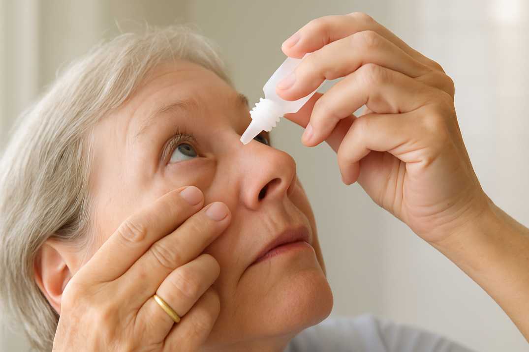
Mastering Eye Drop Administration
Proper technique ensures medication effectiveness and minimizes side effects:
- Wash your hands thoroughly before handling eye drops
- Tilt your head back or lie down for easier application
- Pull down your lower eyelid to create a small pocket
- Hold the bottle upside down just above your eye without touching it to your eye or eyelid
- Squeeze one drop into the pocket (one drop is sufficient)
- Close your eye gently and press lightly on the tear duct at the inner corner for two to three minutes
- Wait at least three to five minutes before applying a second medication if needed
Pro tips for success:
- Link eye drop administration to daily routines like tooth brushing for better adherence
- Set phone alarms or use medication reminder apps
- Keep a chart to track doses, especially helpful when using multiple medications
- Store drops in the refrigerator if you have trouble feeling them land (you’ll feel the cold sensation)
- Bring all medication bottles to appointments so your doctor can verify you’re using the correct medications
- Consider 90-day mail-order prescriptions to avoid running out
Lifestyle Modifications That Make a Difference
Research supports specific dietary patterns for slowing AMD progression:
- Leafy greens (spinach, kale, collards) are rich in lutein and zeaxanthin
- Fatty fish (salmon, mackerel, sardines) provide omega-3 fatty acids
- Citrus fruits and berries are loaded with vitamin C and antioxidants
- Nuts and seeds offer vitamin E
- Colorful vegetables like carrots and sweet potatoes contain vitamin A
The Mediterranean diet pattern shows particular promise for reducing AMD risk and progression.
Physical Activity Benefits
Regular exercise improves blood flow to your eyes and helps manage conditions like diabetes and high blood pressure that increase eye disease risk. Aim for at least 150 minutes of moderate activity weekly, such as brisk walking, swimming, or cycling.
Smoking Cessation
Smoking doubles your risk of developing cataracts and significantly increases AMD risk. Quitting smoking at any age benefits your eyes and overall health. Your healthcare provider can recommend effective cessation programs and medications.
Sun Protection
Ultraviolet (UV) light exposure contributes to both cataract development and AMD. Wear sunglasses with 100% UV protection whenever outdoors, even on cloudy days. Consider wrap-around styles for maximum protection.
Living Well with Vision Changes
Adapting Your Home Environment
Optimize Lighting
Proper lighting makes an enormous difference in daily function:
- Use adjustable lamps with flexible arms to direct light where needed
- Install under-cabinet lighting in the kitchen for safer food preparation
- Add sensor-activated lights in hallways and staircases to prevent falls
- Use LED bulbs labeled “warm” with less blue tone, which work better for people with vision impairment
- Avoid excessive blue light with lampshades that diffuse light and minimize glare
- Keep multiple light sources in each room rather than relying on a single overhead fixture
Increase Contrast
Creating visual contrast helps you identify objects more easily:
- Place dark switch plates or colored tape around light switches and doorknobs
- Arrange brightly colored pillows or throws on the seating to make the furniture visible
- Apply strips of colored tape to step edges to prevent falls
- Use paint or nail polish in bright or dark colors on frequently used items like hairbrushes and kitchen tools
- Choose dishes and placemats in contrasting colors
- Mark oven and microwave controls with raised dots or bright stickers
Leverage Technology
Modern technology offers remarkable solutions:
- Voice assistants (Amazon Echo, Google Home) can set alarms, make calls, read news, and control smart home devices
- Smartphone magnification apps enlarge text and provide text-to-speech capabilities
- Screen readers convert written text to speech on computers and tablets
- Large-button phones and talking watches for easier use
- Electronic magnifiers (CCTV systems) project enlarged images onto screens
- Audiobook services through the National Library Service (free) or commercial platforms
Low Vision Aids and Assistive Devices
Magnification Options
Several magnifier types serve different purposes:
Handheld magnifiers offer portability for reading labels, price tags, and menus while shopping or dining out. Many include built-in lights for better visibility.
Stand magnifiers rest above the page, keeping your hands free for writing or crafting. They’re perfect for prolonged reading sessions and maintain consistent focal distance.
Electronic magnifiers allow adjustable magnification levels and can reverse contrast (white text on black background), often easier for people with AMD to read.
Wearable magnifiers attach to eyeglasses or come as specialized low-vision glasses with telescopic or prismatic lenses.
Reading Modifications
- Select large-print books, newspapers, and magazines with bold fonts
- Use e-readers that allow customizable font sizes and bolding
- Place clear yellow sheets over printed pages to increase contrast
- Adjust computer, tablet, and phone settings for larger text and reverse contrast
- Explore free audiobooks through the National Library Service
Mobility and Independence
Using Your Peripheral Vision
AMD typically spares peripheral vision. Learn to use this remaining vision by:
- Holding brightly colored objects in front of you and moving them up, down, left, and right to find your clearest peripheral area
- Practicing looking “around” the central blind spot by turning or tilting your head
- Enrolling in eccentric viewing training with a low vision specialist
Mobility Aids
- Basic white canes provide support and information about the terrain ahead. Standard white canes typically cost $20 to $60
- Smart canes with GPS and object detection offer enhanced navigation, ranging from $100 to over $1,000, depending on features
- Walkers with larger wheels navigate different surfaces more easily
Consider working with an orientation and mobility specialist who can teach safe navigation techniques specific to your vision loss pattern.
Maintaining Social Connections and Mental Health
Vision loss significantly impacts emotional well-being. Depression and anxiety commonly accompany chronic eye conditions, but resources can help:
Stay socially engaged:
- Join support groups specifically for people with vision loss
- Participate in activities adapted for low vision (audiobook clubs, gentle exercise classes)
- Use video calls with family positioned to maximize your functional vision
- Attend low vision exhibitions and support group meetings to learn about new aids and connect with others
Seek mental health support when needed:
- Talk with your doctor if you feel persistently sad or anxious for more than two weeks
- Consider counseling to process grief about vision changes
- Contact support organizations like the Macular Society (0300 3030 111) or American Macular Degeneration Foundation (888-622-8527)
- Recognize that Charles Bonnet syndrome (visual hallucinations common with vision loss) is a known condition that your doctor can help manage
Coordinating Your Care Team
Building Your Eye Health Partnership
Effective chronic eye condition management requires collaboration between you and various specialists:
Your primary eye care provider (optometrist or ophthalmologist) conducts regular comprehensive exams and monitors disease progression.
Glaucoma specialists focus specifically on glaucoma diagnosis and treatment, offering access to advanced procedures and clinical trials.
Retina specialists manage wet AMD and other retinal conditions, administering anti-VEGF injections and monitoring disease activity.
Low vision specialists evaluate your remaining vision and recommend assistive devices and rehabilitation strategies tailored to your specific needs.
Occupational therapists trained in low vision can visit your home, assess your environment, and suggest practical modifications for safer, more independent living.
Maximizing Your Appointments
Prepare for each visit to get the most value:
- Write down questions beforehand
- Bring all current eye medications in their bottles
- Note any vision changes since your last visit
- Ask about new treatments and clinical trial opportunities
- Request written instructions for any new medications or techniques
- Bring a family member or friend to help remember information discussed
- Understand your target eye pressure numbers (for glaucoma) or monitoring schedule (for AMD)
Insurance Coverage and Financial Resources
Medicare and Medicaid Benefits
Medicare Part B typically covers:
- Annual dilated eye exams for diabetics and high-risk individuals
- Glaucoma screening for high-risk patients
- Diagnostic tests and treatment for eye diseases
- Some low vision rehabilitation services
- One pair of glasses or contact lenses after cataract surgery
Medicaid coverage varies by state but generally includes comprehensive eye exams and necessary treatments. At Poudre Valley Eyecare, we accept both Medicare and Medicaid, making quality eye care accessible to Northern Colorado seniors.
Reducing Out-of-Pocket Costs
Consider these strategies:
- Ask about generic medication options, which cost significantly less than brand names
- Explore patient assistance programs offered by pharmaceutical companies
- Request 90-day supplies through mail order to reduce copays
- Inquire about clinical trials, which provide cutting-edge treatments at no cost
- Check if your county offers senior services programs with vision care assistance
Prevention and Risk Reduction Strategies
Can You Prevent These Conditions?
While you can’t completely prevent age-related eye diseases, you can significantly reduce your risk:
Control chronic health conditions:
- Maintain healthy blood pressure (high blood pressure increases glaucoma and AMD risk)
- Manage diabetes carefully (diabetic retinopathy compounds vision problems)
- Keep cholesterol levels in healthy ranges
Schedule regular eye exams:
- Annual comprehensive exams after age 65 catch problems early
- More frequent exams (every 6-12 months) if you have risk factors
- Never skip appointments, even if your vision seems fine
Know your family history:
- First-degree relatives with glaucoma or AMD increase your risk substantially
- Share this information with your eye doctor for appropriate monitoring
- Consider genetic testing when family history is strong
Protect your eyes from injury:
- Wear protective eyewear during sports and home projects
- Use proper safety equipment at work
- Previous eye injuries increase glaucoma risk
What to Do If Diagnosed: Next Steps
Processing the Diagnosis
Receiving a diagnosis of chronic eye disease can feel overwhelming. Remember:
- Early detection offers the best outcomes
- Treatments can preserve your remaining vision
- Millions of people manage these conditions successfully
- You don’t face this journey alone
Give yourself time to process the information, ask questions, and reach out for support.
Taking Action Immediately
For glaucoma:
- Start prescribed eye drops exactly as directed
- Never skip doses (set multiple reminders if needed)
- Report any side effects to your doctor
- Schedule follow-up appointments as recommended
- Consider joining glaucoma support groups
For AMD:
- Begin AREDS2 supplements if recommended (for intermediate or advanced AMD)
- Schedule anti-VEGF injections promptly if you have wet AMD
- Use your Amsler grid daily to monitor for changes
- Make dietary improvements immediately
- Quit smoking if you currently smoke
- Explore low vision services early rather than waiting
Long-Term Success Strategies
Track your progress:
- Keep a vision journal, noting any changes
- Document medication side effects or problems with adherence
- Record your eye pressure readings and visual field test results
- Note questions between appointments
Stay informed:
- Join glaucoma and AMD organizations for updates on research
- Subscribe to reputable eye health newsletters
- Attend educational seminars offered by eye care centers
- Ask your doctor about clinical trial opportunities
Advocate for yourself:
- Speak up about difficulties with medications or treatments
- Request referrals to specialists when needed
- Ask about accommodations for low vision at work or in public spaces
- Connect with disability services if vision loss impacts daily function
Research Advances: Hope on the Horizon
Exciting Developments in Glaucoma Research
Beyond the gene therapy and iDose TR implant mentioned earlier, several promising areas show potential:
Neuroprotective therapies aim to protect optic nerve cells from damage regardless of eye pressure. Researchers are studying:
- Nicotinamide (vitamin B3) to increase cellular energy and reduce oxidative stress
- Citicoline to reduce retinal ganglion cell loss
- Combination approaches with CoQ10 for enhanced protection
Artificial intelligence is revolutionizing glaucoma detection and monitoring, with algorithms identifying disease progression from visual field tests and OCT images more accurately than traditional methods.
Continuous IOP monitoring using smart contact lenses provides real-time pressure data, helping doctors identify dangerous pressure spikes missed by single office measurements.
AMD Research Breakthroughs
Stem cell therapies are advancing rapidly, with researchers testing whether injected retinal cells can replace damaged ones in dry AMD. Several clinical trials are currently recruiting participants.
Gene therapy approaches similar to those for glaucoma are being adapted for AMD, targeting pathways that lead to cell death and abnormal blood vessel growth.
Novel drug delivery systems, including longer-acting formulations and sustained-release implants, could reduce the burden of frequent anti-VEGF injections for wet AMD.
Scientific Research Supporting This Article
Study 1: Frontiers in Medicine – Glaucoma Treatment Advances (2025)
A comprehensive review published in Frontiers in Medicine examined recent breakthroughs in glaucoma therapeutics, highlighting the approval and efficacy of ROCK inhibitors like ripasudil and netarsudil for lowering intraocular pressure. The research detailed how these medications work through novel mechanisms to enhance fluid outflow from the eye. Additionally, the study covered latanoprostene bunod, a nitric oxide-donating prostaglandin approved by the FDA that demonstrates superior IOP reduction compared to traditional timolol therapy. This systematic review emphasized how these new drug classes offer alternatives for patients who don’t respond adequately to first-line treatments, representing significant progress in glaucoma management.
Source: Sarkis, S., Chamard, C., Johansen, B., Daien, V., & Michon, F. (2025). Challenging glaucoma with emerging therapies. Frontiers in Medicine, 12. https://doi.org/10.3389/fmed.2025.1527319
Study 2: WebMD – Photobiomodulation Therapy for Dry AMD (January 2025)
Groundbreaking research presented at the American Society of Retina Specialists meeting documented the FDA approval of photobiomodulation (PBM) therapy as the first treatment specifically for vision loss from dry AMD. Dr. Eleonora Lad from Duke University Medical Center led clinical trials demonstrating that PBM delivers specific wavelengths of light to the retina, improving cellular function by increasing mitochondrial energy production, decreasing inflammation, and enhancing nutrient delivery to cells. The treatment showed meaningful effects in dry AMD patients and offers hope for intervention at earlier stages before photoreceptor loss becomes irreversible, marking a paradigm shift in dry AMD management after decades without effective treatment options.
Source: WebMD. (2025, January 7). New therapy brings hope for dry AMD vision loss. https://www.webmd.com/eye-health/macular-degeneration/news/20250107/new-therapy-brings-hope-for-dry-amd-vision-loss
Study 3: International Journal of Molecular Sciences – Gene Therapy for Glaucoma (2024)
Scientists at Trinity College Dublin developed an innovative gene therapy targeting matrix metalloproteinase-3 (MMP-3) that demonstrated significant neuroprotective benefits in animal models of glaucoma and human retinal cells derived from glaucoma patients. The research, published in the International Journal of Molecular Sciences, showed that the therapy protected crucial retinal ganglion cells from damage and improved their function. In human retinal cells, the gene therapy increased oxygen consumption and ATP (energy) production, indicating enhanced cellular performance. This represents a potential paradigm shift from merely managing eye pressure to actually protecting and potentially regenerating the optic nerve cells damaged by glaucoma.
Source: Millington-Ward, S., Palfi, A., Shortall, C., Finnegan, L. K., Bargroff, E., Post, I. J. M., Maguire, J., Irnaten, M., O’Brien, C., Kenna, P. F., Chadderton, N., & Farrar, G. J. (2024). AAV-NDI1 therapy provides significant benefit to murine and cellular models of glaucoma. International Journal of Molecular Sciences, 25(16), 8876. https://doi.org/10.3390/ijms25168876
Additional Resources and Citations
For those seeking more detailed information about managing glaucoma and AMD, these authoritative resources provide comprehensive, evidence-based guidance:
Resource 1: CDC Vision and Eye Health Surveillance System (VEHSS)
The Centers for Disease Control and Prevention’s Vision and Eye Health Surveillance System provides the most current national data on glaucoma and AMD prevalence, including state and county-level estimates. Their 2024 reports offer valuable insights into disease patterns and public health strategies.
Access at: https://www.cdc.gov/vision-health-data/
Resource 2: National Eye Institute – Eye Health Information
The National Eye Institute, part of the National Institutes of Health, offers patient-friendly resources on glaucoma and AMD, including symptoms, treatment options, and the latest research developments. Their materials are available in multiple languages and are designed for various reading levels.
Access at: https://www.nei.nih.gov/learn-about-eye-health/eye-conditions-and-diseases
Resource 3: BrightFocus Foundation – Glaucoma and Macular Degeneration Support
BrightFocus Foundation provides free educational materials, webinars, and direct access to experts for people living with glaucoma and macular degeneration. Their resources include practical tips for daily living, medication management guides, and information about clinical trials.
Access at: https://www.brightfocus.org/
Your Vision, Your Future
Managing chronic eye conditions like glaucoma and AMD after 65 requires commitment, but the rewards are substantial. With today’s treatments and the remarkable advances on the horizon, maintaining functional vision throughout your senior years is achievable.
Remember these key takeaways:
- Regular comprehensive eye exams remain your most powerful tool for preserving vision
- Early detection and treatment offer the best outcomes
- Daily medication adherence makes the difference between stable vision and progression
- Lifestyle modifications complement medical treatments effectively
- Low vision aids and adaptations maintain independence even with vision changes
- Support resources help you navigate emotional and practical challenges
- Ongoing research continues to bring new hope for better treatments
At Poudre Valley Eyecare, we’ve spent 25+ years partnering with Northern Colorado families in their eye health journey. We accept Medicare and Medicaid, making comprehensive care accessible to everyone in our community. Whether you need routine monitoring, advanced treatment, or low vision services, we’re here to provide the expert, compassionate care you deserve.
Don’t let chronic eye conditions steal your independence or quality of life. Schedule your comprehensive eye exam today and take the first step toward preserving your vision for years to come. Your future self will thank you for the investment you make in your eye health now.
FAQs
-
-
Take prescribed eye drops daily, attend regular eye exams, and discuss laser or surgical options with an eye doctor for optimal control.
-
Please note: None of the above should be considered medical advice. If you’re having any concerns about your vision, please reach out to us immediately or see your primary care provider.


