
What Is Retinal Imaging and Do You Need It? A Clear Explanation
Introduction
If you’ve been to an eye doctor recently, you may have been offered retinal imaging for an additional cost of $25-60. Most eye care practices charge between $25 to $60 for this service, with the typical range being $35-50, and vision plans generally do not cover the cost of it. This leaves many patients wondering: what exactly is retinal imaging, and is it worth the extra expense?
Bottom Line Up Front: Retinal imaging is a valuable diagnostic tool that creates detailed digital pictures of the back of your eye, helping detect serious eye diseases and systemic health conditions early—often before symptoms appear. While not always necessary for everyone, it’s generally worth the cost for comprehensive eye health monitoring and can serve as an excellent complement to traditional eye exams.
What Is Retinal Imaging?
Retinal imaging takes digital pictures of the back of your eye, including the part that light and images hit, called the retina. Think of it as a high-resolution camera specifically designed to photograph the inside of your eye, capturing detailed images of your retina, optic disc, optic nerve, and blood vessels.
At Poudre Valley Eyecare, we use advanced retinal imaging technology to provide comprehensive eye health assessments for our Fort Collins community.
How Retinal Imaging Works
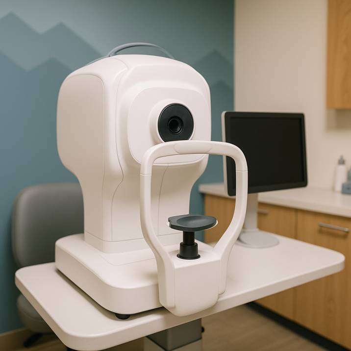
The process is surprisingly simple and completely painless:
- Positioning: You sit comfortably and place your chin on a rest while looking into the imaging device
- Targeting: Your doctor will direct you to look into the machine one eye at a time. It might seem a bit like looking through a peephole.
- Capturing: When you see a brief flash of light, you’ll know the image has been taken.n
- Review: The images from the test should be ready immediately. Normally, your doctor will talk to you about them before you leave.
Types of Retinal Imaging Technology
Fundus Photography: Color and black-and-white photography that creates detailed images of your retinal surface.
Optical Coherence Tomography (OCT): A scanner uses light waves to form an image of the retina on a computer screen, providing cross-sectional views of retinal layers.
Ultra-Widefield Imaging: Advanced systems like the Optomap laser retina scan can capture 82% of your retina at once, compared to traditional imaging techniques that display only 15% of your retina at one time.
Who Needs Retinal Imaging?
High-Risk Individuals
Retinal imaging is particularly beneficial for individuals at high risk for retinal diseases. Your eye doctor may recommend the test if you have diabetes, suspected retinal toxicity, macular degeneration, or glaucoma.
You should strongly consider retinal imaging if you have:
- Diabetes or Pre-diabetes: Even early-stage diabetes can cause retinal damage before other symptoms appear
- Family History of Eye Diseases: Genetic predisposition to glaucoma, macular degeneration, or other retinal conditions increases your risk significantly
- Hypertension: Signs of high blood pressure, diabetes, and some forms of cancer are first evident in the retina, often well before other signs appear throughout the body
- Age Over 60: Substantially increased risk for age-related eye conditions
- Previous Eye Injuries or Surgeries: Ongoing monitoring for potential complications
Everyone Else: The Preventive Care Approach
Even if you’re not in a high-risk category, retinal imaging offers significant value for preventive care. For most patients, weighing the pros and cons of retinal imaging, the investment is worthwhile when considering both immediate and long-term health benefits.
Benefits of Retinal Imaging
Early Disease Detection
These images can be used as a diagnostic tool to help detect vision- or life-threatening disorders and diseases early, often before symptoms appear. Early detection is crucial because many serious eye conditions cause irreversible damage if caught too late.
Conditions that retinal imaging can detect:
- Diabetic retinopathy
- Glaucoma (early optic nerve damage)
- Age-related macular degeneration
- Retinal detachment
- Eye tumors
- Stroke indicators
- Cardiovascular disease signs
Comprehensive Health Monitoring
Your retina provides the only location in your body where blood vessels can be directly visualized non-invasively. By examining your retina during an eye exam, your eye doctor can identify signs of eye disease as well as indicators of other health problems, including stroke, heart disease, diabetes, and hypertension.
Historical Documentation and Longitudinal Comparison
Retinal imaging creates a permanent digital record of your eye’s interior structures, enabling your doctor to compare images over time and track subtle changes that might indicate developing problems. This historical baseline is invaluable for monitoring treatment outcomes and detecting disease progression in its earliest stages.
Retinal Imaging vs. Eye Dilation: Understanding the Difference
Many patients assume retinal imaging replaces the need for eye dilation, but this isn’t entirely accurate.
What Eye Dilation Offers
Traditional dilation uses drops to widen your pupils, allowing your doctor to examine your entire retina manually. Eye dilation can make it hard to see up close for 1-2 hours and make your eyes light-sensitive for 4-6 hours.
Retinal Imaging Limitations
While retinal imaging can reduce the likelihood of requiring eye dilation, it has important limitations. Even advanced “ultra-widefield” retinal imaging captures only approximately 80% of the retinal surface, leaving the peripheral areas unexamined.
When Dilation Is Still Necessary
Therefore, eye dilation is still necessary in specific situations to view the edges of the retina. For example, if you have a sudden increase in spots in vision, flashes of light, or a dark curtain coming across your vision, a dilated eye examination is the gold standard to detect any retinal breaks.
Cost and Insurance Coverage: What You Need to Know
Typical Costs
According to current 2024-2025 pricing data, the cost of retinal imaging depends on several factors, including the specific imaging technology used, your geographic location, and your insurance coverage. Most practices charge between $35-40 per session, though costs can range from $25 to $60 depending on the provider and technology used.
Insurance Coverage Reality
It is considered “healthy eye imaging,” which is not covered under most vision plans. On the other hand, dilation is considered diagnostic imaging when completed as part of a routine eye exam.
Important coverage notes:
- Vision insurance: Typically doesn’t cover retinal imaging
- Medical insurance: Retinal imaging may be covered by your medical insurance (not your vision insurance) or Medicare. It depends on the terms of your police, as well as the reason you are having the test done.
- Medicare: May cover when medically necessary
Cost-Benefit Analysis
When considering the expense, it’s important to weigh the cost of preventive screening against potential treatment costs for advanced eye disease. Early detection through retinal imaging can prevent more expensive interventions later and preserve irreplaceable vision.
Recent Scientific Advances in Retinal Imaging
Breakthrough Research Findings
According to the CDC, in 2021, around 9.6 million people of all ages were living with diabetic retinopathy in the U.S. Among them, around 1.84 million had vision-threatening diabetic retinopathy. The retinal imaging devices market has grown significantly, reaching USD 5.79 billion in 2024 and is expected to continue growing at a CAGR of 7.8% through 2030. In a new study published January 24th in Science Translational Medicine, physician-researchers from Mass Eye and Ear, a member of Mass General Brigham, and the Broad Institute of MIT and Harvard combined retinal imaging, genetic, and big data to estimate how likely a person is to develop eye and systemic diseases in the future.
Artificial Intelligence Integration
The integration of artificial intelligence represents a significant advancement in retinal imaging capabilities. In April 2024, AEYE-DS became the first fully autonomous AI algorithm to receive FDA clearance for diagnosing referable diabetic retinopathy from retinal images taken by handheld cameras. This breakthrough technology achieved sensitivity rates of 92-93% and specificity rates of 89-94% in large-scale clinical trials.
Predictive Health Capabilities
Research has identified significant associations between retinal layer thinning and increased risk of developing ocular, cardiac, pulmonary, metabolic, and neuropsychiatric diseases. Scientists have also identified specific genes associated with retinal layer thickness, opening new possibilities for personalized medicine approaches.
Making the Decision: Should You Get Retinal Imaging?
When Retinal Imaging Is Highly Recommended
- You have diabetes or pre-diabetes
- Family history of eye diseases exists
- You’re over 60 years old
- You have hypertension or cardiovascular disease
- You’ve experienced recent vision changes
- Your eye care provider specifically recommends it based on risk assessment
When It May Be Optional
- You’re under 40 with no significant risk factors
- You’ve had recent retinal imaging with normal results
- Budget constraints are significant, and no risk factors are present
Questions to Discuss with Your Eye Care Provider
- “Based on my health history and risk factors, do you recommend retinal imaging?”
- “What specific conditions are you screening for in my particular case?”
- “How frequently should I have this examination performed?”
- “Will my insurance plan cover any portion of this service?”
- “What would you personally do if you were in my situation?”
What to Expect During Your Retinal Imaging Appointment
Preparation Requirements
- No special preparation needed: Unlike dilation procedures, retinal imaging typically requires no preparation.
- Bring sunglasses: You may experience temporary light sensitivity and blurred vision if dilation is also performed.
- Transportation planning: Avoid driving, screen use, or reading immediately after the procedure if your pupils are dilated
During the Imaging Procedure
- Quick and painless: The entire imaging process takes just a few minutes
- Non-contact procedure: Nothing touches your eye during the examination
- Immediate results: Images are available for review immediately following capture
Post-Imaging Consultation
- Results discussion: Your doctor will review the images with you in detail
- Follow-up planning: Determine the appropriate timing for your next screening
- Educational opportunity: This is an ideal time to understand what your images reveal about your eye health
Conclusion
Retinal imaging represents a significant advancement in preventive eye care, offering early detection capabilities that can preserve vision and identify life-threatening systemic diseases. While the out-of-pocket investment may seem substantial, the long-term health benefits—particularly for individuals with risk factors—generally justify the expense.
Key Clinical Takeaways:
- Retinal imaging provides detailed visualization of ocular structures, detecting problems before symptoms manifest
- The technology is particularly valuable for patients with diabetes, a family history of eye disease, or those over 60
- The procedure is rapid, painless, and provides immediate diagnostic results
- While typically not covered by vision insurance, medical insurance may provide coverage when clinically indicated
- Recent AI advances are significantly improving diagnostic accuracy and accessibility
The clinical decision ultimately depends on your risk profile, financial considerations, and personal health priorities. When uncertain, consultation with your eye care provider can offer personalized guidance based on your specific health history and current ocular status.
Remember, vision loss is often irreversible. Early detection and preventive intervention are consistently more effective and cost-efficient than treating advanced pathology. At Poudre Valley Eyecare, we remain committed to helping patients make informed decisions about their ocular health, supported by over 25 years of clinical experience serving the Fort Collins community.
For more information about comprehensive eye exams or diabetic eye care, explore our other educational resources.
The Future of Retinal Imaging
Emerging Technologies
Recent advances in retinal imaging technology include fluorescence lifetime imaging ophthalmoscopy, which can reveal biochemical and metabolic changes at the cellular level. Optical coherence tomography continues to evolve toward higher resolution, faster imaging speeds, increased portability, and improved cost-effectiveness.
Enhanced Accessibility
Portable devices and telemedicine integration are significantly expanding access to retinal imaging, particularly benefiting underserved populations and remote geographic areas.
Additional Resources and Citations
For those interested in learning more about retinal imaging research and advances, here are key scientific sources referenced in this article:
-
Mass Eye and Ear Predictive Health Research (2024)
“Retinal imaging and genetics data used to predict future disease risk”
Published in Science Translational Medicine, this groundbreaking study demonstrates how retinal imaging combined with genetic analysis can predict future disease risk across multiple health conditions.
Read the full study -
Advances in AI for Diabetic Retinopathy Detection (2024)
“Advances in Structural and Functional Retinal Imaging and Biomarkers for Early Detection of Diabetic Retinopathy”
This comprehensive review in PMC explores the latest developments in multimodal imaging and AI algorithms, including the FDA-approved AEYE-DS system, achieving 92-93% sensitivity.
Access the research -
Retinal Imaging Market Analysis and Technology Trends (2025)
“Retinal Imaging Devices Market Size & Share Report”
Industry analysis showing the retinal imaging market reaching $5.79 billion in 2024, with 7.8% annual growth driven by AI integration and increased accessibility.
View market insights
Schedule your comprehensive eye examination today to discuss whether retinal imaging is appropriate for your clinical situation. Our experienced clinical team accepts Medicare and Medicaid, ensuring quality eye care remains accessible to all community members.
Call us or schedule online to begin your retinal health assessment.
FAQs
-
Retinal imaging is a painless, non-invasive test where a camera captures detailed pictures of the back of your eye to spot early signs of disease or damage
Please note: None of the above should be considered medical advice. If you’re having any concerns about your vision, please reach out to us immediately or see your primary care provider.

